New study challenges theory about deep-sea fish mystery
The researchers examined the retinas of fish larvae caught at deep depths
For over a century, biology textbooks have asserted that vertebrate vision, including our own, is constructed from two distinct cell types: rods for processing dim light and cones for bright light and colour.
However, groundbreaking new research involving deep-sea fish is now challenging this long-held understanding.
Scientists have identified a novel type of visual cell in these deep-sea creatures that remarkably blends the physical form of rods with the molecular machinery and genes typically associated with cones.
This hybrid cell, uniquely adapted for sight in the perpetually gloomy conditions of the deep ocean, was discovered in the larval stages of three distinct deep-sea fish species inhabiting the Red Sea.
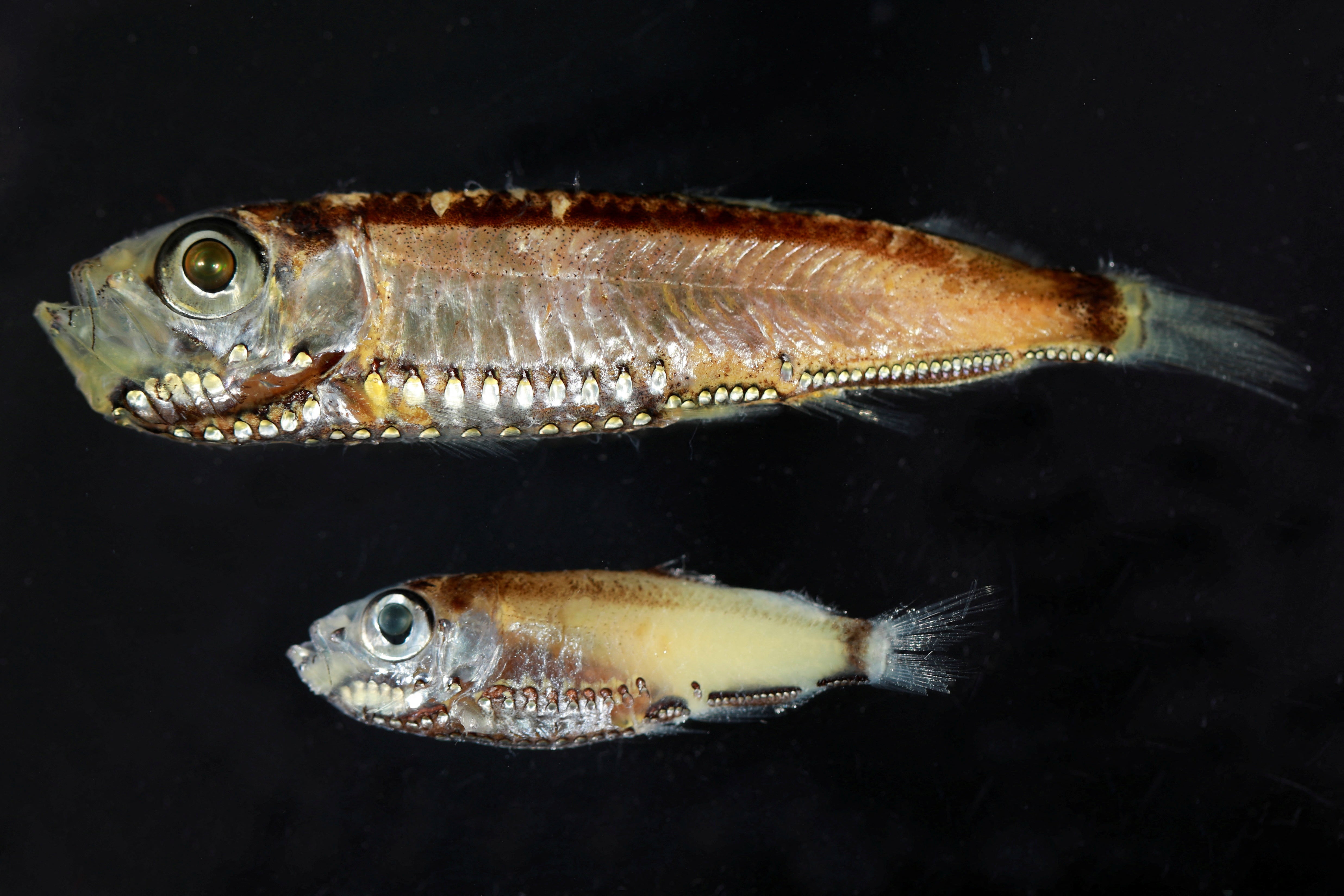
The species studied included a hatchetfish (Maurolicus mucronatus), a lightfish (Vinciguerria mabahiss), and a lanternfish (Benthosema pterotum).
While the hatchetfish retained these hybrid cells throughout its life, the other two species transitioned to the conventional rod-cone dichotomy as adults.
These small fish, with adults typically measuring between 3-7 cm, and their larvae even smaller, thrive in a marine realm where sunlight struggles to penetrate the watery depths.
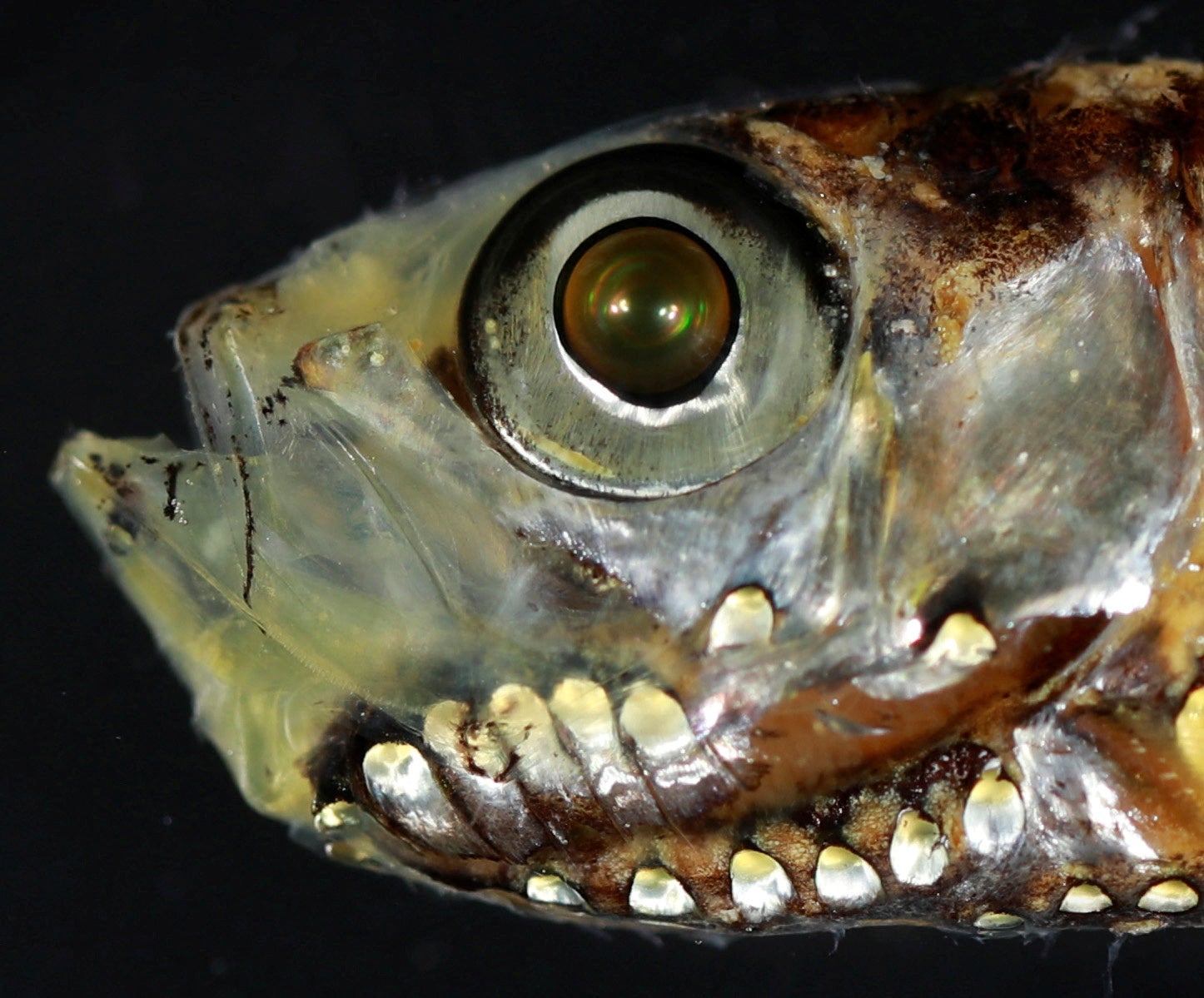
The vertebrate retina, the sensory membrane at the back of the eye responsible for detecting light and converting it into signals for the brain, traditionally possesses two main types of light-sensitive visual cells, known as photoreceptors, named for their characteristic rod and cone shapes.
"The rods and cones slowly change position inside the retina when moving between dim and bright conditions, which is why our eyes take time to adjust when we flick on the light switch on our way to the restroom at night," said Lily Fogg, a postdoctoral researcher in marine biology at the University of Helsinki in Finland and lead author of the research published in the journal Science Advances.
"We found that, as larvae, these deep-sea fish mostly use a mix-and-match type of hybrid photoreceptor. These cells look like rods - long, cylindrical and optimized to catch as many light particles - photons - as possible. But they use the molecular machinery of cones, switching on genes usually found only in cones," Fogg said.
The researchers examined the retinas of fish larvae caught at depths from 65 to 650 feet (20 to 200 meters).
In the type of dim environment they inhabit, rod and cone cells both are usually engaged in the vertebrate retina, but neither works very well. These fish display an evolutionary remedy.
"Our results challenge the longstanding idea that rods and cones are two fixed, clearly separated cell types. Instead, we show that photoreceptors can blend structural and molecular features in unexpected ways. This suggests that vertebrate visual systems are more flexible and evolutionarily adaptable than previously thought," Fogg said.
"It is a very cool finding that shows that biology does not fit neatly into boxes," said study senior author Fabio Cortesi, a marine biologist and neuroscientist at the University of Queensland in Australia. "I wouldn't be surprised if we find these cells are much more common across all vertebrates, including terrestrial species."
All three species emit bioluminescence using small light-emitting organs on their bodies, mostly located on the belly. They produce blue-green light that blends with the faint background light from the sun above. This strategy, called counterillumination, is a common form of camouflage in the deep sea to avoid predators.
"Small fish like these fuel the open ocean. They are plentiful and serve as food for many larger predatory fishes, including tuna and marlin, marine mammals such as dolphins and whales, and marine birds," Cortesi said.
These kinds of fish also engage in one of the biggest daily migrations in the animal kingdom. They swim near the surface at night to feed in plankton-rich waters, then return to the depths - 650 to 3,280 feet (200 to 1,000 meters) - during daytime to avoid predation.
"The deep sea remains a frontier for human exploration, a mystery box with the potential for significant discoveries," Cortesi said. "We should look after this habitat with the utmost care to make sure future generations can continue to marvel at its wonders."
Join our commenting forum
Join thought-provoking conversations, follow other Independent readers and see their replies
Comments
Bookmark popover
Removed from bookmarks